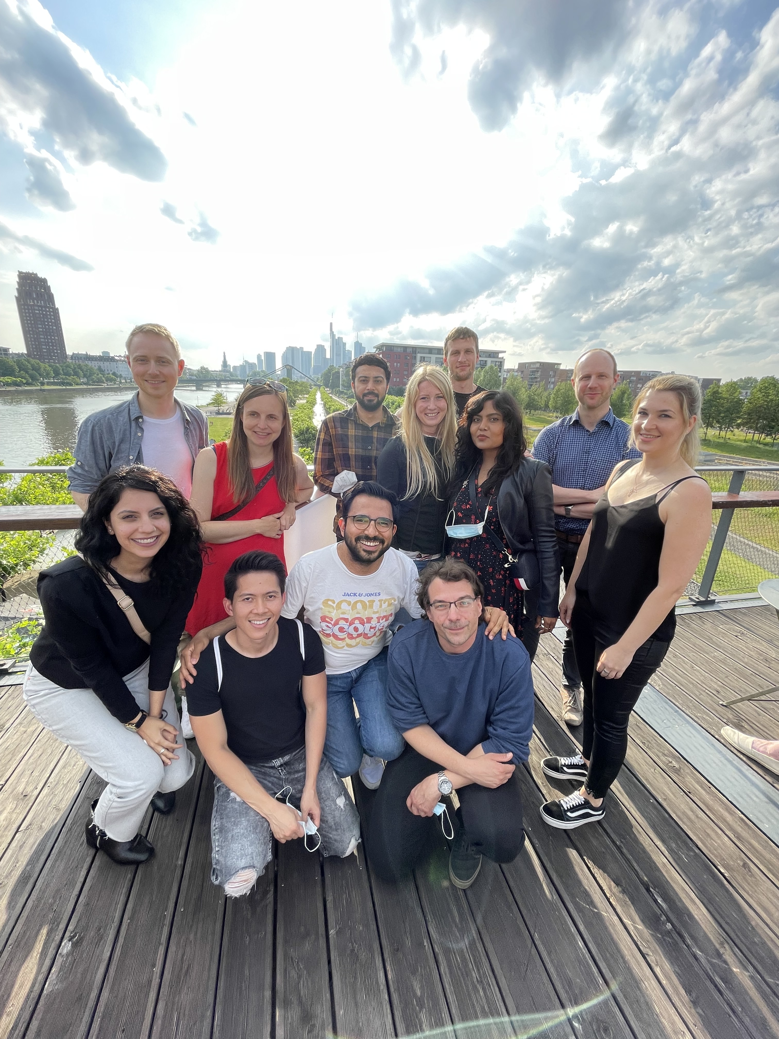
Welcome to the
Münch lab
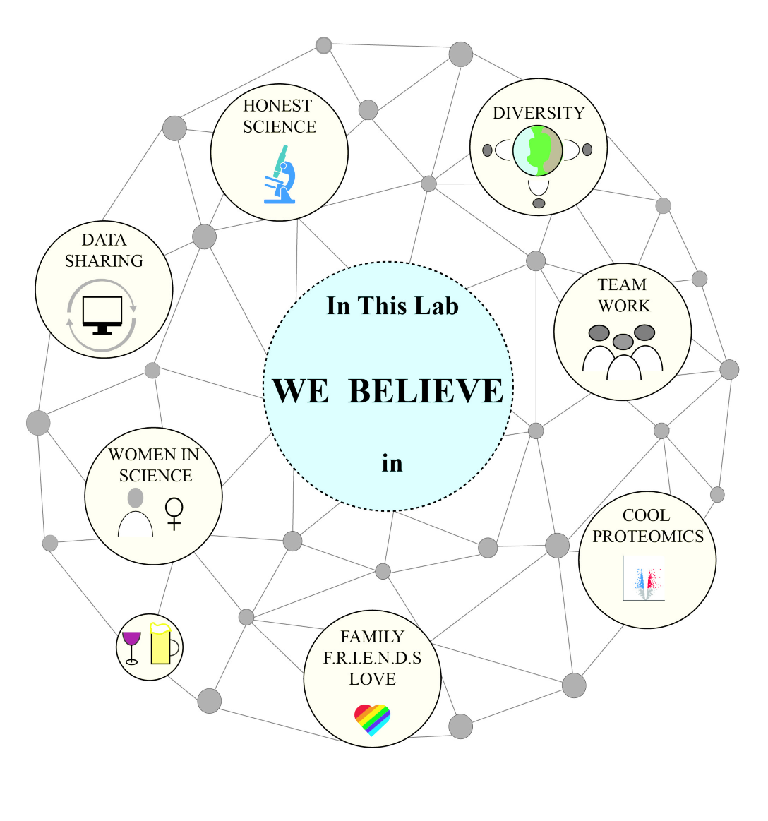
We are a international, multidisciplinary team focusing on the molecular mechanisms of life. Applying a combination of systems biology, biochemistry and molecular biology, we examine how stress responses shape cellular behavior and pathology. Our team consists of biochemists, physicians, pharmacists, bioinformaticians and biologists. Based in the collaborative environment of Frankfurt we strive to discover the big picture of biology.
Our Research
To find out more click on one of the main topics or directly explore the detailed projects:


Selected Publications
(first/last author publications)
Show All PublicationsAll Publications
Publications from PubMed: 98
Zhou X, Kuenne C, Günther S, Wittig I, Güven B, Boutguetait D, Delgado Lagos F, Sachs N, Maegdefessel L, Müller OJ, Münch C, Offermanns S, Fleming I, Siragusa M Nuclear eNOS Interacts With and -Nitrosates ADAR1 to Modulate Type I Interferon Signaling and Endothelial Function. Circulation 2025. Link
Gotthardt G, Weckesser J, Tascher G, Barros da Gama S, Uckelmann HJ, Sun S, Schwalm MP, Mosler T, Ferrario G, Friedmann Angeli JP, Münch C, Knapp S, Müller S Cysteine-reactive covalent chloro-N-acetamide ligands induce ferroptosis mediated cell death. EMBO Rep 2025. Link
Esparza-Moltó PB, Goswami AV, Bozkurt S, Münch C, Newman LE, Moyzis AG, Rojas GR, Guan D, Jones JR, Gage FH, Shadel GS ROS-dependent localization of glycolytic enzymes to mitochondria. Redox Biol 2025. 86 103812 Link
Lopez M, Herrle N, Amirmiran B, Malacarne PF, Werkhäuser J, Chatterjee S, Kader C, Jurisch V, Wen X, Gheisari M, Schäfer K, Münch C, Leuschner F, Gilsbach R, Rezende F, Brandes RP Prolonged In Vivo Chemogenetic Generation of Hydrogen Peroxide by Endothelial Cells Induces Cardiac Remodelling and Vascular Dysfunction. Antioxidants (Basel) 2025. 14 (6) Link
Mukherjee R, Bhattacharya A, Tomaskovic I, Mello-Vieira J, Brunstein ME, Başoğlu M, Veenendaal T, Bailey H, Colby T, Misra M, Eimer S, Klumperman J, Münch C, Matic I, Dikic I Phosphoribosyl ubiquitination of SNARE proteins regulates autophagy during Legionella infection. EMBO J 2025. 44 (15) 4252-4279 Link
Lorenz NI, Sauer B, Urban H, Weinem JB, Parmar BS, Zeiner PS, Strecker MI, Schulte D, Mittelbronn M, Alekseeva T, Sevenich L, Harter PN, Münch C, Steinbach JP, Luger AL, Heiland DH, Ronellenfitsch MW AMP-activated protein kinase mediates adaptation of glioblastoma cells to conditions of the tumor microenvironment. J Exp Clin Cancer Res 2025. 44 (1) 104 Link
Moser LM, Heim C, Koschade SE, Wendel P, Bozkurt S, Harenkamp S, Kreyenberg H, Merker M, Münch C, Gradhand E, Vogler M, Ullrich E, Bönig H, Klusmann JH, Bader P, Wels WS, Rettinger E CAR-CIK vs. CAR-T: benchmarking novel cytokine-induced killer cells as solid tumor immunotherapy in ErbB2+ rhabdomyosarcoma. Front Immunol 2025. 16 1485817 Link
Sauer B, Kueckelhaus J, Lorenz NI, Bozkurt S, Schulte D, Weinem JB, Benzarti M, Meiser J, Urban H, Villa G, Harter PN, Münch C, Rieger J, Steinbach JP, Heiland DH, Ronellenfitsch MW An AMP-activated protein kinase-PGC-1α axis mediates metabolic plasticity in glioblastoma. Clin Transl Med 2024. 14 (11) e70030 Link
Prieto-Garcia C, Matkovic V, Mosler T, Li C, Liang J, Oo JA, Haidle F, Mačinković I, Cabrera-Orefice A, Berkane R, Giuliani G, Xu F, Jacomin AC, Tomaskovic I, Basoglu M, Hoffmann ME, Rathore R, Cetin R, Boutguetait D, Bozkurt S, Hernández Cañás MC, Keller M, Busam J, Shah VJ, Wittig I, et al. Pathogenic proteotoxicity of cryptic splicing is alleviated by ubiquitination and ER-phagy. Science 2024. 386 (6723) 768-776 Link
Foged MM, Recazens E, Chollet S, Lisci M, Allen GE, Zinshteyn B, Boutguetait D, Münch C, Mootha VK, Jourdain AA Cytosolic dependent translation supports mitochondrial RNA processing. Proc Natl Acad Sci U S A 2024. 121 (47) e2414187121 Link
Bozkurt S, Parmar BS, Münch C Quantifying mitochondrial protein import by mePROD proteomics. Methods Enzymol 2024. 706 449-474 Link
Sanz-Martinez P, Tascher G, Cano-Franco S, Cabrerizo-Poveda P, Münch C, Homan EJ, Stolz A GPCR Function in Autophagy Control: A Systematic Approach of Chemical Intervention. J Mol Biol 2024. 436 (15) 168643 Link
Eickhorst C, Babic R, Rush-Kittle J, Lucya L, Imam FL, Sánchez-Martín P, Hollenstein DM, Michaelis J, Münch C, Meisinger C, Slade D, Gámez-Díaz L, Kraft C FIP200 Phosphorylation Regulates Late Steps in Mitophagy. J Mol Biol 2024. 436 (15) 168631 Link
Verheyden NA, Klostermann M, Brüggemann M, Steede HM, Scholz A, Amr S, Lichtenthaeler C, Münch C, Schmid T, Zarnack K, Krueger A A high-resolution map of functional miR-181 response elements in the thymus reveals the role of coding sequence targeting and an alternative seed match. Nucleic Acids Res 2024. 52 (14) 8515-8533 Link
Koch B, Shehata M, Müller-Ruttloff C, Gouda SA, Wetzstein N, Patyna S, Scholz A, Schmid T, Dietrich U, Münch C, Ziebuhr J, Geiger H, Martinez-Sobrido L, Baer PC, Mostafa A, Pleschka S Influenza A virus replicates productively in primary human kidney cells and induces factors and mechanisms related to regulated cell death and renal pathology observed in virus-infected patients. Front Cell Infect Microbiol 2024. 14 1363407 Link
Brenner D, Sieverding K, Srinidhi J, Zellner S, Secker C, Yilmaz R, Dyckow J, Amr S, Ponomarenko A, Tunaboylu E, Douahem Y, Schlag JS, Rodríguez Martínez L, Kislinger G, Niemann C, Nalbach K, Ruf WP, Uhl J, Hollenbeck J, Schirmer L, Catanese A, Lobsiger CS, Danzer KM, Yilmazer-Hanke D, Münch C, et al. A TBK1 variant causes autophagolysosomal and motoneuron pathology without neuroinflammation in mice. J Exp Med 2024. 221 (5) Link
Sauer B, Lorenz NI, Divé I, Klann K, Luger AL, Urban H, Schröder JH, Steinbach JP, Münch C, Ronellenfitsch MW Mammalian target of rapamycin inhibition protects glioma cells from temozolomide-induced cell death. Cell Death Discov 2024. 10 (1) 8 Link
Kuntschar S, Cardamone G, Klann K, Bauer R, Meyer SP, Raue R, Rappl P, Münch C, Brüne B, Schmid T Mmp12 Is Translationally Regulated in Macrophages during the Course of Inflammation. Int J Mol Sci 2023. 24 (23) Link
Mende H, Khatri A, Lange C, Poveda-Cuevas SA, Tascher G, Covarrubias-Pinto A, Löhr F, Koschade SE, Dikic I, Münch C, Bremm A, Brunetti L, Brandts CH, Uckelmann H, Dötsch V, Rogov VV, Bhaskara RM, Müller S An atypical GABARAP binding module drives the pro-autophagic potential of the AML-associated NPM1c variant. Cell Rep 2023. 42 (12) 113484 Link
Rehbein S, Possmayer AL, Bozkurt S, Lotsch C, Gerstmeier J, Burger M, Momma S, Maletzki C, Classen CF, Freiman TM, Dubinski D, Lamszus K, Stringer BW, Herold-Mende C, Münch C, Kögel D, Linder B Molecular Determinants of Calcitriol Signaling and Sensitivity in Glioma Stem-like Cells. Cancers (Basel) 2023. 15 (21) Link
Reisbeck L, Linder B, Tascher G, Bozkurt S, Weber KJ, Herold-Mende C, van Wijk SJL, Marschalek R, Schaefer L, Münch C, Kögel D The iron chelator and OXPHOS inhibitor VLX600 induces mitophagy and an autophagy-dependent type of cell death in glioblastoma cells. Am J Physiol Cell Physiol 2023. 325 (6) C1451-C1469 Link
Pereira RS, Kumar R, Cais A, Paulini L, Kahler A, Bravo J, Minciacchi VR, Krack T, Kowarz E, Zanetti C, Godavarthy PS, Hoeller F, Llavona P, Stark T, Tascher G, Nowak D, Meduri E, Huntly BJP, Münch C, Pampaloni F, Marschalek R, Krause DS Distinct and targetable role of calcium-sensing receptor in leukaemia. Nat Commun 2023. 14 (1) 6242 Link
Lambert M, Gebel J, Trejtnar C, Wesch N, Bozkurt S, Adrian-Allgood M, Löhr F, Münch C, Dötsch V Fuzzy interactions between the auto-phosphorylated C-terminus and the kinase domain of CK1δ inhibits activation of TAp63α. Sci Rep 2023. 13 (1) 16423 Link
Münch C, Kirstein J Protein quality control: from molecular mechanisms to therapeutic intervention-EMBO workshop, May 21-26 2023, Srebreno, Croatia. Cell Stress Chaperones 2023. 28 (6) 631-640 Link
Brat C, Huynh Phuoc HP, Awad O, Parmar BS, Hellmuth N, Heinicke U, Amr S, Grimmer J, Sürün D, Husnjak K, Carlsson M, Fahrer J, Bauer T, Krieg SC, Manolikakes G, Zacharowski K, Steinhilber D, Münch C, Maier TJ, Roos J Endogenous anti-tumorigenic nitro-fatty acids inhibit the ubiquitin-proteasome system by directly targeting the 26S proteasome. Cell Chem Biol 2023. 30 (10) 1277-1294.e12 Link
Sutandy FXR, Gößner I, Tascher G, Münch C A cytosolic surveillance mechanism activates the mitochondrial UPR. Nature 2023. 618 (7966) 849-854 Link
Maier CR, Hartmann O, Prieto-Garcia C, Al-Shami KM, Schlicker L, Vogel FCE, Haid S, Klann K, Buck V, Münch C, Schmitz W, Einig E, Krenz B, Calzado MA, Eilers M, Popov N, Rosenfeldt MT, Diefenbacher ME, Schulze A USP28 controls SREBP2 and the mevalonate pathway to drive tumour growth in squamous cancer. Cell Death Differ 2023. 30 (7) 1710-1725 Link
Bhattacharya A, Mukherjee R, Kuncha SK, Brunstein ME, Rathore R, Junek S, Münch C, Dikic I A lysosome membrane regeneration pathway depends on TBC1D15 and autophagic lysosomal reformation proteins. Nat Cell Biol 2023. 25 (5) 685-698 Link
Metzler M, Tharyan RG, Klann K, Grikscheit K, Bojkova D, Cinatl J, Tascher G, Ciesek S, Münch C SARS-CoV-2 Variants Show Different Host Cell Proteome Profiles With Delayed Immune Response Activation in Omicron-Infected Cells. Mol Cell Proteomics 2023. 22 (5) 100537 Link
Schäfer JA, Sutandy FXR, Münch C Omics-based approaches for the systematic profiling of mitochondrial biology. Mol Cell 2023. 83 (6) 911-926 Link
Sánchez-Martín P, Kriegenburg F, Alves L, Adam J, Elsaesser J, Babic R, Mancilla H, Licheva M, Tascher G, Münch C, Eimer S, Kraft C ULK1-mediated phosphorylation regulates the conserved role of YKT6 in autophagy. J Cell Sci 2023. 136 (3) Link
Räuber S, Schroeter CB, Strippel C, Nelke C, Ruland T, Dik A, Golombeck KS, Regner-Nelke L, Paunovic M, Esser D, Münch C, Rosenow F, van Duijn M, Henes A, Ruck T, Amit I, Leypoldt F, Titulaer MJ, Wiendl H, Meuth SG, Meyer Zu Hörste G, Melzer N Cerebrospinal fluid proteomics indicates immune dysregulation and neuronal dysfunction in antibody associated autoimmune encephalitis. J Autoimmun 2023. 135 102985 Link
Michaelis JB, Bozkurt S, Schäfer JA, Münch C Monitoring Mitochondrial Protein Import Using Mitochondrial Targeting Sequence (MTS)-eGFP. Bio Protoc 2022. 12 (24) Link
Nasrullah U, Stanke K, Recknagel V, Bozkurt S, Wurzel P, Gauer S, Imre G, Münch C, Pfeilschifter J, Eberhardt W The E3 Ligase TRIM25 Impairs Apoptotic Cell Death in Colon Carcinoma Cells via Destabilization of Caspase-7 mRNA: A Possible Role of hnRNPH1. Cells 2023. 12 (1) Link
Key J, Gispert S, Koornneef L, Sleddens-Linkels E, Kohli A, Torres-Odio S, Koepf G, Amr S, Reichlmeir M, Harter PN, West AP, Münch C, Baarends WM, Auburger G CLPP Depletion Causes Diplotene Arrest; Underlying Testis Mitochondrial Dysfunction Occurs with Accumulation of Perrault Proteins ERAL1, PEO1, and HARS2. Cells 2022. 12 (1) Link
Walther M, Vestweber PK, Kühn S, Rieger U, Schäfer J, Münch C, Vogel-Kindgen S, Planz V, Windbergs M Bioactive Insulin-Loaded Electrospun Wound Dressings for Localized Drug Delivery and Stimulation of Protein Expression Associated with Wound Healing. Mol Pharm 2023. 20 (1) 241-254 Link
Merline R, Rödig H, Zeng-Brouwers J, Poluzzi C, Tascher G, Michaelis J, Lopez-Mosqueda J, Rhiner A, Huber LS, Diehl V, Dikic I, Kögel D, Münch C, Wygrecka M, Schaefer L A20 binding and inhibitor of nuclear factor kappa B (NF-κB)-1 (ABIN-1): a novel modulator of mitochondrial autophagy. Am J Physiol Cell Physiol 2023. 324 (2) C339-C352 Link
Schmitt D, Bozkurt S, Henning-Domres P, Huesmann H, Eimer S, Bindila L, Behrends C, Boyle E, Wilfling F, Tascher G, Münch C, Behl C, Kern A FACS-mediated isolation of native autophagic vesicles. Autophagy 2023. 19 (7) 2146-2147 Link
Wu Z, Berlemann LA, Bader V, Sehr DA, Dawin E, Covallero A, Meschede J, Angersbach L, Showkat C, Michaelis JB, Münch C, Rieger B, Namgaladze D, Herrera MG, Fiesel FC, Springer W, Mendes M, Stepien J, Barkovits K, Marcus K, Sickmann A, Dittmar G, Busch KB, Riedel D, Brini M, et al. LUBAC assembles a ubiquitin signaling platform at mitochondria for signal amplification and transport of NF-κB to the nucleus. EMBO J 2022. 41 (24) e112006 Link
Koschade SE, Klann K, Shaid S, Vick B, Stratmann JA, Thölken M, Meyer LM, Nguyen TD, Campe J, Moser LM, Hock S, Baker F, Meyer CT, Wempe F, Serve H, Ullrich E, Jeremias I, Münch C, Brandts CH Correction: Translatome proteomics identifies autophagy as a resistance mechanism to on-target FLT3 inhibitors in acute myeloid leukemia. Leukemia 2022. 36 (12) 2950 Link
Koschade SE, Tascher G, Parmar BS, Brandts CH, Münch C SpinTip: A Simple, Robust, and Versatile Preanalytical Method for Microscale Suspension Cell Proteomics. J Proteome Res 2022. 21 (11) 2827-2835 Link
Schmitt D, Bozkurt S, Henning-Domres P, Huesmann H, Eimer S, Bindila L, Behrends C, Boyle E, Wilfling F, Tascher G, Münch C, Behl C, Kern A Lipid and protein content profiling of isolated native autophagic vesicles. EMBO Rep 2022. 23 (12) e53065 Link
Divé I, Klann K, Michaelis JB, Heinzen D, Steinbach JP, Münch C, Ronellenfitsch MW Inhibition of mTOR signaling protects human glioma cells from hypoxia-induced cell death in an autophagy-independent manner. Cell Death Discov 2022. 8 (1) 409 Link
Michaelis JB, Brunstein ME, Bozkurt S, Alves L, Wegner M, Kaulich M, Pohl C, Münch C Protein import motor complex reacts to mitochondrial misfolding by reducing protein import and activating mitophagy. Nat Commun 2022. 13 (1) 5164 Link
Karantanou C, Minciacchi VR, Kumar R, Zanetti C, Bravo J, Pereira RS, Tascher G, Tertel T, Covarrubias-Pinto A, Bankov K, Pfeffermann LM, Bonig H, Divieti-Pajevic P, McEwan DG, Giebel B, Münch C, Dikic I, Krause DS Impact of mesenchymal stromal cell-derived vesicular cargo on B-cell acute lymphoblastic leukemia progression. Blood Adv 2023. 7 (7) 1190-1203 Link
Koschade SE, Klann K, Shaid S, Vick B, Stratmann JA, Thölken M, Meyer LM, Nguyen TD, Campe J, Moser LM, Hock S, Baker F, Meyer CT, Wempe F, Serve H, Ullrich E, Jeremias I, Münch C, Brandts CH Translatome proteomics identifies autophagy as a resistance mechanism to on-target FLT3 inhibitors in acute myeloid leukemia. Leukemia 2022. 36 (10) 2396-2407 Link
Sutandy FXR, Tharyan RG, Münch C The beginning of a new era: pioneering direct screens for RNA modulators. Signal Transduct Target Ther 2022. 7 (1) 246 Link
Delaunay S, Pascual G, Feng B, Klann K, Behm M, Hotz-Wagenblatt A, Richter K, Zaoui K, Herpel E, Münch C, Dietmann S, Hess J, Benitah SA, Frye M Mitochondrial RNA modifications shape metabolic plasticity in metastasis. Nature 2022. 607 (7919) 593-603 Link
Fischer T, Hartmann O, Reissland M, Prieto-Garcia C, Klann K, Pahor N, Schülein-Völk C, Baluapuri A, Polat B, Abazari A, Gerhard-Hartmann E, Kopp HG, Essmann F, Rosenfeldt M, Münch C, Flentje M, Diefenbacher ME PTEN mutant non-small cell lung cancer require ATM to suppress pro-apoptotic signalling and evade radiotherapy. Cell Biosci 2022. 12 (1) 50 Link
Prieto-Garcia C, Hartmann O, Reissland M, Braun F, Bozkurt S, Pahor N, Fuss C, Schirbel A, Schülein-Völk C, Buchberger A, Calzado Canale MA, Rosenfeldt M, Dikic I, Münch C, Diefenbacher ME USP28 enables oncogenic transformation of respiratory cells, and its inhibition potentiates molecular therapy targeting mutant EGFR, BRAF and PI3K. Mol Oncol 2022. 16 (17) 3082-3106 Link
Klann K, Münch C Quantitative Translation Proteomics Using mePROD. Methods Mol Biol 2022. 2428 75-87 Link
Klann K, Münch C PBLMM: Peptide-based linear mixed models for differential expression analysis of shotgun proteomics data. J Cell Biochem 2022. 123 (3) 691-696 Link
Kohli A, Sauerhering L, Fehling SK, Klann K, Geiger H, Becker S, Koch B, Baer PC, Strecker T, Münch C Proteomic landscape of SARS-CoV-2- and MERS-CoV-infected primary human renal epithelial cells. Life Sci Alliance 2022. 5 (5) Link
Li X, Straub J, Medeiros TC, Mehra C, den Brave F, Peker E, Atanassov I, Stillger K, Michaelis JB, Burbridge E, Adrain C, Münch C, Riemer J, Becker T, Pernas LF Mitochondria shed their outer membrane in response to infection-induced stress. Science 2022. 375 (6577) eabi4343 Link
Schäfer JA, Bozkurt S, Michaelis JB, Klann K, Münch C Global mitochondrial protein import proteomics reveal distinct regulation by translation and translocation machinery. Mol Cell 2022. 82 (2) 435-446.e7 Link
Prieto-Garcia C, Hartmann O, Reissland M, Fischer T, Maier CR, Rosenfeldt M, Schülein-Völk C, Klann K, Kalb R, Dikic I, Münch C, Diefenbacher ME Inhibition of USP28 overcomes Cisplatin-resistance of squamous tumors by suppression of the Fanconi anemia pathway. Cell Death Differ 2022. 29 (3) 568-584 Link
Gama-Brambila RA, Chen J, Dabiri Y, Tascher G, Němec V, Münch C, Song G, Knapp S, Cheng X A Chemical Toolbox for Labeling and Degrading Engineered Cas Proteins. JACS Au 2021. 1 (6) 777-785 Link
Engel AL, Lorenz NI, Klann K, Münch C, Depner C, Steinbach JP, Ronellenfitsch MW, Luger AL Correction: Serine-dependent redox homeostasis regulates glioblastoma cell survival. Br J Cancer 2021. 125 (7) 1030 Link
Gerstmeier J, Possmayer AL, Bozkurt S, Hoffmann ME, Dikic I, Herold-Mende C, Burger MC, Münch C, Kögel D, Linder B Calcitriol Promotes Differentiation of Glioma Stem-Like Cells and Increases Their Susceptibility to Temozolomide. Cancers (Basel) 2021. 13 (14) Link
Lorenz NI, Sittig ACM, Urban H, Luger AL, Engel AL, Münch C, Steinbach JP, Ronellenfitsch MW Activating transcription factor 4 mediates adaptation of human glioblastoma cells to hypoxia and temozolomide. Sci Rep 2021. 11 (1) 14161 Link
Gama-Brambila RA, Chen J, Zhou J, Tascher G, Münch C, Cheng X A PROTAC targets splicing factor 3B1. Cell Chem Biol 2021. 28 (11) 1616-1627.e8 Link
Hahn M, Covarrubias-Pinto A, Herhaus L, Satpathy S, Klann K, Boyle KB, Münch C, Rajalingam K, Randow F, Choudhary C, Dikic I SIK2 orchestrates actin-dependent host response upon infection. Proc Natl Acad Sci U S A 2021. 118 (19) Link
Klann K, Tascher G, Münch C Virus systems biology: Proteomics profiling of dynamic protein networks during infection. Adv Virus Res 2021. 109 1-29 Link
Geßner A, Koch B, Klann K, Fuhrmann DC, Farmand S, Schubert R, Münch C, Geiger H, Baer PC Characterization of Extracellular Vesicles from Preconditioned Human Adipose-Derived Stromal/Stem Cells. Int J Mol Sci 2021. 22 (6) Link
Schwich OD, Blümel N, Keller M, Wegener M, Setty ST, Brunstein ME, Poser I, Mozos IRL, Suess B, Münch C, McNicoll F, Zarnack K, Müller-McNicoll M SRSF3 and SRSF7 modulate 3'UTR length through suppression or activation of proximal polyadenylation sites and regulation of CFIm levels. Genome Biol 2021. 22 (1) 82 Link
Klionsky DJ, Abdel-Aziz AK, Abdelfatah S, Abdellatif M, Abdoli A, Abel S, Abeliovich H, Abildgaard MH, Abudu YP, Acevedo-Arozena A, Adamopoulos IE, Adeli K, Adolph TE, Adornetto A, Aflaki E, Agam G, Agarwal A, Aggarwal BB, Agnello M, Agostinis P, Agrewala JN, Agrotis A, Aguilar PV, Ahmad ST, Ahmed ZM, et al. Guidelines for the use and interpretation of assays for monitoring autophagy (4th edition). Autophagy 2021. 17 (1) 1-382 Link
Klann K, Münch C [Not Available]. Biospektrum (Heidelb) 2021. 27 (1) 40-45 Link
Meyer N, Henkel L, Linder B, Zielke S, Tascher G, Trautmann S, Geisslinger G, Münch C, Fulda S, Tegeder I, Kögel D Autophagy activation, lipotoxicity and lysosomal membrane permeabilization synergize to promote pimozide- and loperamide-induced glioma cell death. Autophagy 2021. 17 (11) 3424-3443 Link
Güllülü Ö, Hehlgans S, Mayer BE, Gößner I, Petraki C, Hoffmann M, Dombrowsky MJ, Kunzmann P, Hamacher K, Strebhardt K, Fokas E, Rödel C, Münch C, Rödel F A Spatial and Functional Interaction of a Heterotetramer Survivin-DNA-PKcs Complex in DNA Damage Response. Cancer Res 2021. 81 (9) 2304-2317 Link
Hiebel C, Stürner E, Hoffmeister M, Tascher G, Schwarz M, Nagel H, Behrends C, Münch C, Behl C BAG3 Proteomic Signature under Proteostasis Stress. Cells 2020. 9 (11) Link
Bojkova D, Bechtel M, McLaughlin KM, McGreig JE, Klann K, Bellinghausen C, Rohde G, Jonigk D, Braubach P, Ciesek S, Münch C, Wass MN, Michaelis M, Cinatl J Aprotinin Inhibits SARS-CoV-2 Replication. Cells 2020. 9 (11) Link
Klann K, Münch C Unbiased translation proteomics upon cell stress. Mol Cell Oncol 2020. 7 (4) 1763150 Link
Klann K, Bojkova D, Tascher G, Ciesek S, Münch C, Cinatl J Growth Factor Receptor Signaling Inhibition Prevents SARS-CoV-2 Replication. Mol Cell 2020. 80 (1) 164-174.e4 Link
McLaughlin KM, Bechtel M, Bojkova D, Münch C, Ciesek S, Wass MN, Michaelis M, Cinatl J COVID-19-Related Coagulopathy-Is Transferrin a Missing Link? Diagnostics (Basel) 2020. 10 (8) Link
Osthues T, Zimmer B, Rimola V, Klann K, Schilling K, Mathoor P, Angioni C, Weigert A, Geisslinger G, Münch C, Scholich K, Sisignano M The Lipid Receptor G2A (GPR132) Mediates Macrophage Migration in Nerve Injury-Induced Neuropathic Pain. Cells 2020. 9 (7) Link
Klann K, Münch C Instrument Logic Increases Identifications during Mutliplexed Translatome Measurements. Anal Chem 2020. 92 (12) 8041-8045 Link
Bojkova D, Klann K, Koch B, Widera M, Krause D, Ciesek S, Cinatl J, Münch C Proteomics of SARS-CoV-2-infected host cells reveals therapy targets. Nature 2020. 583 (7816) 469-472 Link
Key J, Maletzko A, Kohli A, Gispert S, Torres-Odio S, Wittig I, Heidler J, Bárcena C, López-Otín C, Lei Y, West AP, Münch C, Auburger G Loss of mitochondrial ClpP, Lonp1, and Tfam triggers transcriptional induction of Rnf213, a susceptibility factor for moyamoya disease. Neurogenetics 2020. 21 (3) 187-203 Link
Engel AL, Lorenz NI, Klann K, Münch C, Depner C, Steinbach JP, Ronellenfitsch MW, Luger AL Serine-dependent redox homeostasis regulates glioblastoma cell survival. Br J Cancer 2020. 122 (9) 1391-1398 Link
Prieto-Garcia C, Hartmann O, Reissland M, Braun F, Fischer T, Walz S, Schülein-Völk C, Eilers U, Ade CP, Calzado MA, Orian A, Maric HM, Münch C, Rosenfeldt M, Eilers M, Diefenbacher ME Maintaining protein stability of ∆Np63 via USP28 is required by squamous cancer cells. EMBO Mol Med 2020. 12 (4) e11101 Link
Klann K, Tascher G, Münch C Functional Translatome Proteomics Reveal Converging and Dose-Dependent Regulation by mTORC1 and eIF2α. Mol Cell 2020. 77 (4) 913-925.e4 Link
Pitzius S, Osterburg C, Gebel J, Tascher G, Schäfer B, Zhou H, Münch C, Dötsch V TA*p63 and GTAp63 achieve tighter transcriptional regulation in quality control by converting an inhibitory element into an additional transactivation domain. Cell Death Dis 2019. 10 (10) 686 Link
Poluzzi C, Nastase MV, Zeng-Brouwers J, Roedig H, Hsieh LT, Michaelis JB, Buhl EM, Rezende F, Manavski Y, Bleich A, Boor P, Brandes RP, Pfeilschifter J, Stelzer EHK, Münch C, Dikic I, Brandts C, Iozzo RV, Wygrecka M, Schaefer L Biglycan evokes autophagy in macrophages via a novel CD44/Toll-like receptor 4 signaling axis in ischemia/reperfusion injury. Kidney Int 2019. 95 (3) 540-562 Link
Nguyen TD, Shaid S, Vakhrusheva O, Koschade SE, Klann K, Thölken M, Baker F, Zhang J, Oellerich T, Sürün D, Derlet A, Haberbosch I, Eimer S, Osiewacz HD, Behrends C, Münch C, Dikic I, Brandts CH Loss of the selective autophagy receptor p62 impairs murine myeloid leukemia progression and mitophagy. Blood 2019. 133 (2) 168-179 Link
Münch C The different axes of the mammalian mitochondrial unfolded protein response. BMC Biol 2018. 16 (1) 81 Link
Meyer N, Zielke S, Michaelis JB, Linder B, Warnsmann V, Rakel S, Osiewacz HD, Fulda S, Mittelbronn M, Münch C, Behrends C, Kögel D AT 101 induces early mitochondrial dysfunction and HMOX1 (heme oxygenase 1) to trigger mitophagic cell death in glioma cells. Autophagy 2018. 14 (10) 1693-1709 Link
Münch C, Dikic I Publisher Correction: Hitchhiking on selective autophagy. Nat Cell Biol 2018. 20 (8) 990 Link
Münch C, Dikic I Hitchhiking on selective autophagy. Nat Cell Biol 2018. 20 (2) 122-124 Link
Yamano K, Wang C, Sarraf SA, Münch C, Kikuchi R, Noda NN, Hizukuri Y, Kanemaki MT, Harper W, Tanaka K, Matsuda N, Youle RJ Endosomal Rab cycles regulate Parkin-mediated mitophagy. Elife 2018. 7 Link
Drané P, Brault ME, Cui G, Meghani K, Chaubey S, Detappe A, Parnandi N, He Y, Zheng XF, Botuyan MV, Kalousi A, Yewdell WT, Münch C, Harper JW, Chaudhuri J, Soutoglou E, Mer G, Chowdhury D TIRR regulates 53BP1 by masking its histone methyl-lysine binding function. Nature 2017. 543 (7644) 211-216 Link
Yang W, Nagasawa K, Münch C, Xu Y, Satterstrom K, Jeong S, Hayes SD, Jedrychowski MP, Vyas FS, Zaganjor E, Guarani V, Ringel AE, Gygi SP, Harper JW, Haigis MC Mitochondrial Sirtuin Network Reveals Dynamic SIRT3-Dependent Deacetylation in Response to Membrane Depolarization. Cell 2016. 167 (4) 985-1000.e21 Link
Münch C, Harper JW Mitochondrial unfolded protein response controls matrix pre-RNA processing and translation. Nature 2016. 534 (7609) 710-3 Link
Ordureau A, Münch C, Harper JW Quantifying ubiquitin signaling. Mol Cell 2015. 58 (4) 660-76 Link
Suraweera A, Münch C, Hanssum A, Bertolotti A Failure of amino acid homeostasis causes cell death following proteasome inhibition. Mol Cell 2012. 48 (2) 242-53 Link
Münch C, Bertolotti A Propagation of the prion phenomenon: beyond the seeding principle. J Mol Biol 2012. 421 (4-5) 491-8 Link
Münch C, Bertolotti A Self-propagation and transmission of misfolded mutant SOD1: prion or prion-like phenomenon? Cell Cycle 2011. 10 (11) 1711 Link
Münch C, O'Brien J, Bertolotti A Prion-like propagation of mutant superoxide dismutase-1 misfolding in neuronal cells. Proc Natl Acad Sci U S A 2011. 108 (9) 3548-53 Link
Münch C, Bertolotti A Exposure of hydrophobic surfaces initiates aggregation of diverse ALS-causing superoxide dismutase-1 mutants. J Mol Biol 2010. 399 (3) 512-25 Link
People


Our Funding
European Research Council
Emmy Noether-Programm der DFG
Bundesministerium für Bildung und Forschung

Else Kröner Fresenius Stiftung

LOEWE Exzellenzinitative des Landes Hessen
SFB1177 - Autophagy
LOEWE Zentrum für Zell- und Gentherapie Frankfurt
Cardio Pulmonary Institute Frankfurt

ENABLE Frankfurt
Proxidrugs
Aventis Foundation
Johanna Quandt Young Academy
EMBO Young Investigator Programme

Fraunhofer Innovation Center

SFB1531